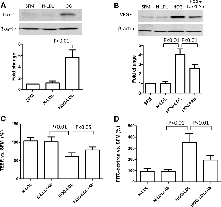
FIG. 4.
LOX-1 was involved in HOG-LDL-induced RPE barrier dysfunction. (A, B) ARPE-19 cells were cultured in 6-well plates for 48 h to reach 80% confluence, and then kept quiescent in SFM for 18 h. Cells were pretreated with versus without a, LOX-1 blocking antibody (50 mg/L, 1 h), and then exposed to N- or HOG-LDL treatment (200 mg/L, 12 h); protein levels of LOX-1 and VEGF were determined by Western blotting and densitometry. (C, D) Experiments were also conducted in the monolayer ARPE-19 barrier model in transwells to determine permeability using TEER and FITC-dextran leakage assays. Data are presented as percentages versus SFM (mean ± SD, n = 3 or 5).

