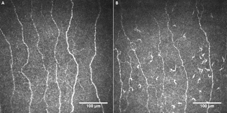
Figure 3.
Representative corneal confocal microscopic images of the subbasal nerve plexus in a patient with multiple sclerosis at baseline (A) and after 2 years (B) showing reduced nerve fibers and increased dendritic cells. This patient had a relapse and an increase of 1.0 point in EDSS score during follow-up.

