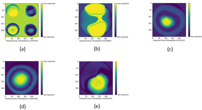
Figure 6.
Selected examples of CAMs showing the areas of an image that are most important for its CNN classification. Classes: 0, keratoconus; 1, normal; 2, subclinical keratoconus. CAMs of a four-map display image (A), a front elevation map (B), a back elevation map (C), a corneal pachymetry map (D), and a front sagittal curvature map (E).

