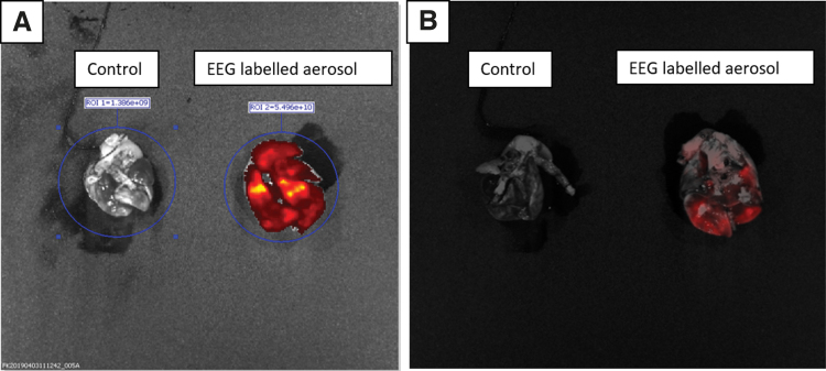
FIG. 3.
Fluorescent images of EEG powder deposition (10 mg). (A) Fluorescence image for control and EEG Survanta powder-labeled aerosol and (B) 3D overlay of the fluorescence with light image mixed for control and EEG Survanta powder-labeled aerosol. Image mixing shows Quasi-complete deposition of EEG Survanta in the distal lung. 3D, three-dimensional. Color images are available online.

