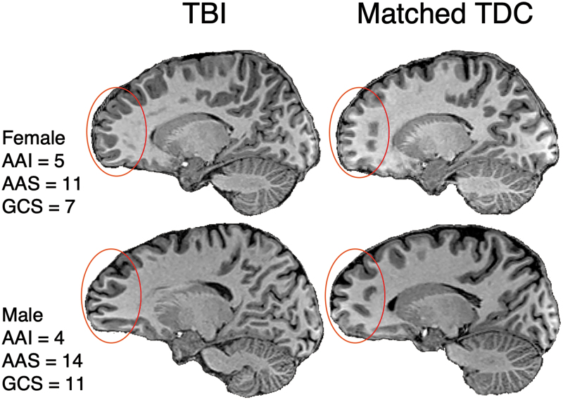
FIG. 2.
Examples of gyrification patterns in two adolescents several years after pediatric TBI compared with two demographically-matched adolescents in the TDC comparison group on conventional T1-weighted imaging. Note the irregular pattern and seemingly deeper gyrification in the medial aspects of the frontal lobes (regions within the overlaid red circle) in the children with TBI versus their age-, sex-, and SES-matched controls. TBI, traumatic brain injury; TDC, typically developing child; AAI, age at injury; AAS, age at scan; GCS, Glasgow Coma Scale score. Color image is available online.

