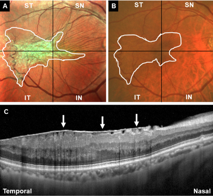
Figure 2.

Representative case on how to have preoperative decision making of ERM demarcation in the MC-SLO group (A) and the CFP group (B). The residents and experienced surgeons were required to have delineation of the whole ERM margin based on the MC-SLO and CFP images (white lines in A and B). (C) OCT image required when having MC-SLO imaging supported the definite diagnosis of ERM (white arrows) in the patient.
