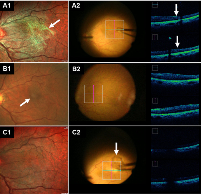
Figure 4.

Representative case on how to have intraoperative agreement assessment in the MC-SLO group (A) and the CFP group (B). All the fundus photographs (A1, B1, and C1) have been rotated 180° to appear the same with the surgeons’ viewing under microscope when performing the ERM surgery. (A) In the MC-SLO group, the location to have initial peeling has been pointed out preoperatively (white arrow in A1), iOCT imaging revealed the selected location was true, as there was more remarkable and obvious elevation of the retina's inner surface (white arrows in A2). (B) In the CFP group, the location to have peeling has been pointed out preoperatively (white arrows in B1), iOCT imaging revealed the selected location was false, as there was no remarkable and obvious elevation of the retina's inner surface. (C) MC-SLO image taken 1 day postoperatively (C1) and iOCT imaging taken intraoperatively (C2) revealed complete peeling off of the ERM (arrows in C2).
