
Figure 6.
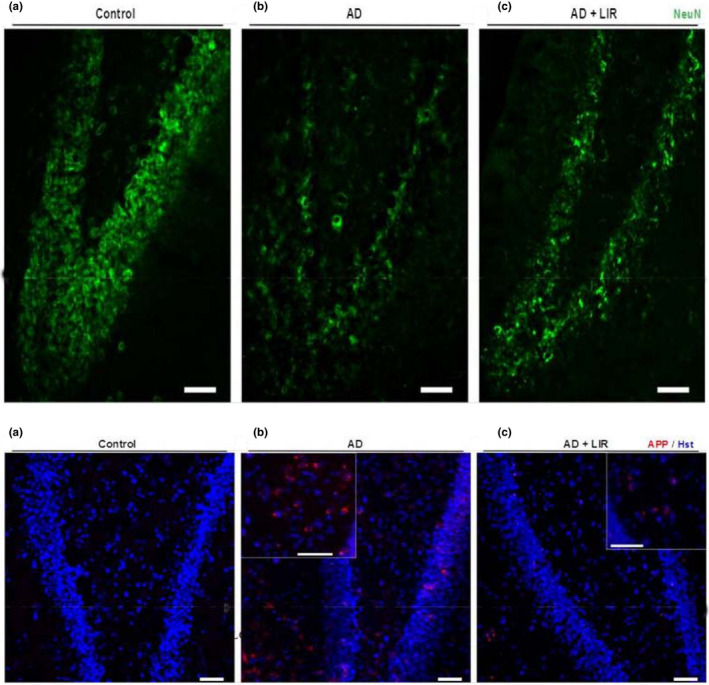
(1): Representative Immunofluorescent images of rat hippocampus immunostained for Anti‐Amyloid Precursor Protein (APP). (a) Coronal section of the albino rat shows negative staining for APP. (b) Apparent deposition of APP in the AD group. (c) AD + LIR group shows a sparse deposition of APP. (Insets) Higher magnification of APP deposits (Scale bar = 50 µm). Figure (6; 2): Representative Immunofluorescent images of rat hippocampus immunostained for Neuronal nuclei (Anti‐NeuN). (a) Coronal section of albino rat shows numerous NeuN‐positive cells in the hippocampal proper. (b) AD group exhibits negative NeuN staining. (c) AD + LIR group restores some positive NeuN cells (Scale bar = 50 µm). (AD = Alzheimer's disease, LIR = liraglutide and LIR + AD = liraglutide +Alzheimer)