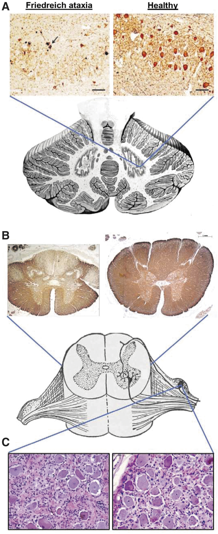
Figure 1.
Key neuropathological features of Friedreich ataxia. (A) Dentate nucleus stained for neurons (neuron-specific enolase), showing loss of large glutaminergic neurons. The arrow indicates surviving small neurons. Reproduced with permission from Koeppen AH et al.(B) Spinal cord cross-sections stained for myelin (myelin basic protein), showing pronounced lack of myelin in the dorsal columns, dorsal spinocerebellar, and corticospinal tracts. (C) Dorsal root ganglion stained with hematoxylin and eosin (nuclei in dark purple and cytoplasm in pink). A reduction in the average size and number of neurons (large cells) and proliferation of satellite cells and monocytes (other nuclei) is evident.

