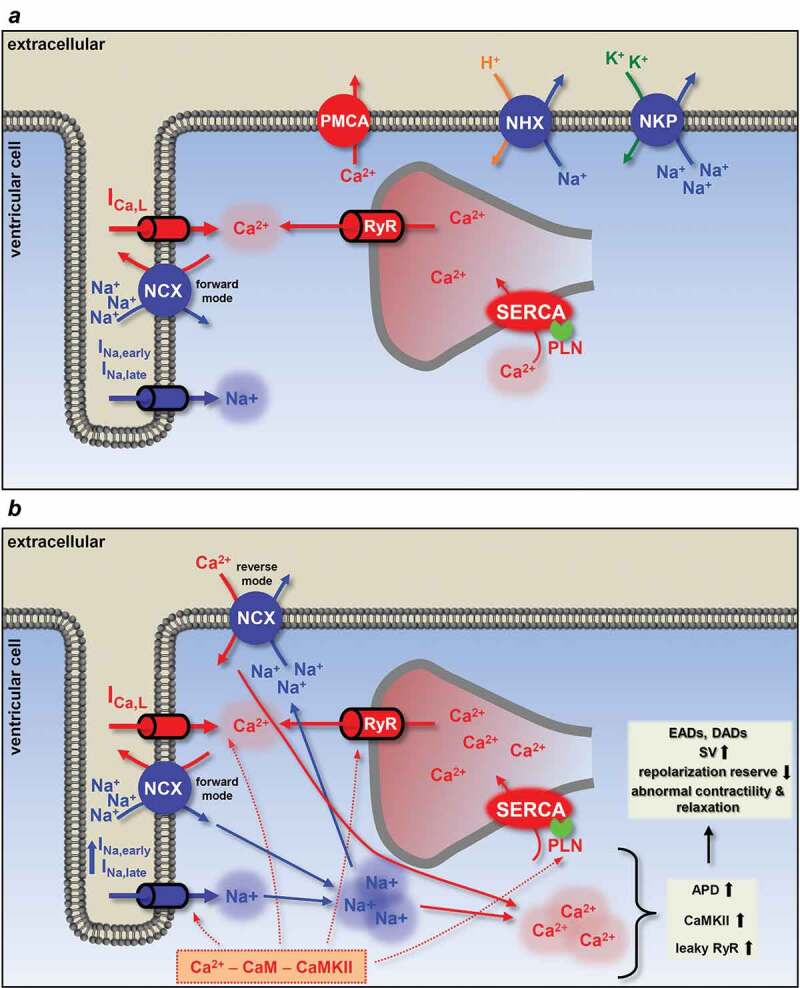
Figure 5.

Schematic illustration of the physiological and pathophysiological processes leading to arrhythmias upon increased INa,late. (a) In a healthy myocyte, excitation-contraction coupling controls contraction by periodically increasing and decreasing the intracellular Ca2+ concentration. (b) If INa,late is elevated, as in the case of many diseases, intracellular Na+ and a concomitant Ca2+ overload may lead to arrhythmias. High intracellular Na+ concentration can activate the reverse mode NCX to further load the cell with Ca2+. Ca2+ overload and the longer action potential duration predispose the cell to proarrhythmic events. Red arrows show Ca2+ related, while blue arrows show Na+ related processes. Dashed lines indicate the phosphorylation targets of the Ca2+ – CaM – CaMKII pathway. APD, action potential duration; CaM, calmodulin; CaMKII, Ca/calmodulin-dependent protein kinase II; DAD, delayed afterdepolarization; EAD, early afterdepolarization; ICa,L, L-type Ca2+ current; INa,early, the fast, early component of the Na+ current; INa,late, the persistent, late component of the Na+ current; NCX, Na+/Ca2+ exchange; NHX, Na+/H+ exchanger; NKP, Na+/K+ pump; PLN, phospholamban; PMCA, plasma membrane Ca2+-ATPase; RyR, ryanodine receptor; SERCA, sarcoplasmic reticulum Ca2+-ATPase; SV, short term beat-to-beat variability of action potential duration
