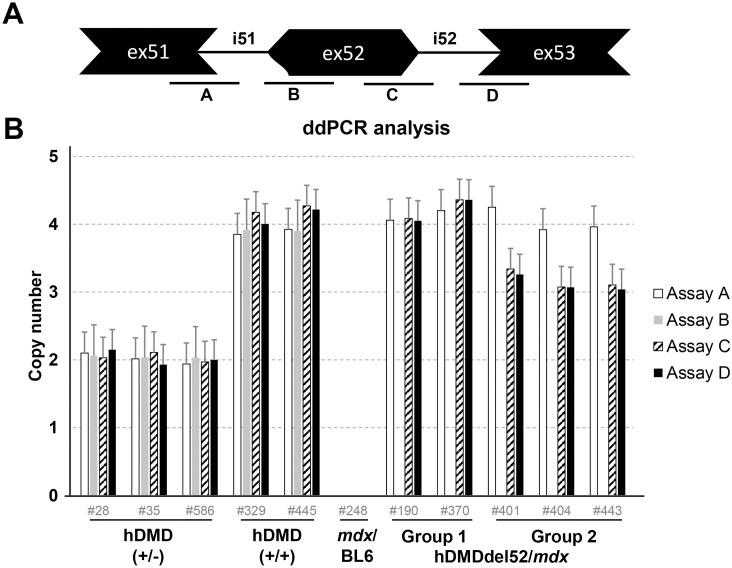
Fig 4. ddPCR copy number analysis.
A. Schematic representation of the probe locations. B. ddPCR analysis of copy numbers of exons 51, 52 and 53 in hDMDdel52/mdx and hDMDdel52-53/mdx mice and controls, relative to Tfrc copy number. Most assays showed a double signal for both hDMD/mdx and hDMDdel52/mdx mice, while no signal was detected in mdx/BL6 mice. For the hDMDdel52/mdx and hDMDdel52-53/mdx mice, the probe on the intron 51- exon 52 boundary (assay B) did not give a signal. However, the probe on the exon 52 –intron 52 boundary did (assay C). As previously observed, assay C and assay D showed lower signals in hDMDdel52-53/mdx mice compared to hDMDdel52/mdx.

