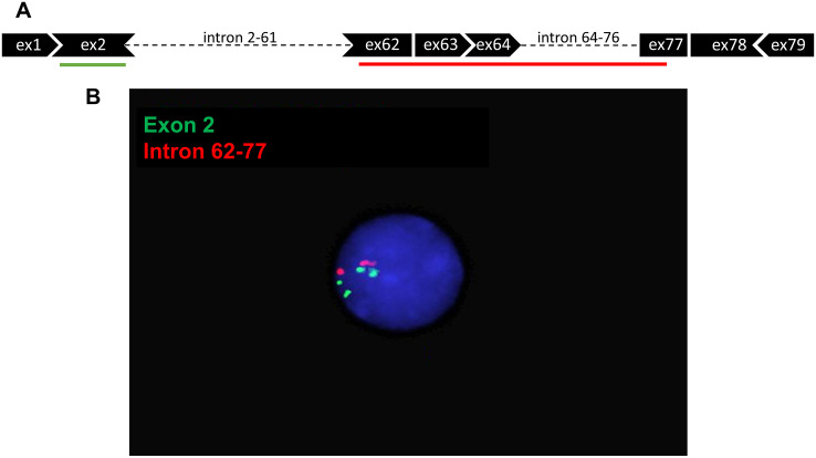
Fig 5. Detection of hDMD tandem duplication in hDMDdel52/mdx mouse by FISH analysis in interphase nuclei.
A. Schematic representation of FISH probes. B. Hybridization results under fluorescent microscopy. Two signals can be seen for the red probe which targets intron 62–77, while four signals are detected for the green probe targeting exon 2. Because metaphase spreads could not be detected, interphase nuclei were used for the analysis. hDMDdel52-53/mdx mice were not included in this analysis.

