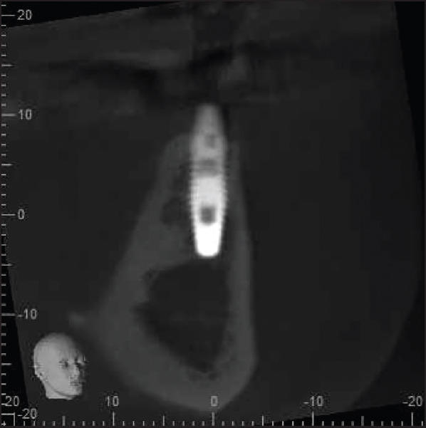
Figure 5.

Coronal section of the CBCT scan shows the implant housed entirely within the alveolar bone. There was a buccal concavity in the bone from the resorption pattern of the alveolar ridge; thus, the implant was placed relatively closer to the buccal bone (but still entirely within the ridge) to enable prosthetically driven placement and future ease of restoration of the implant
CBCT: cone-beam computed tomography
