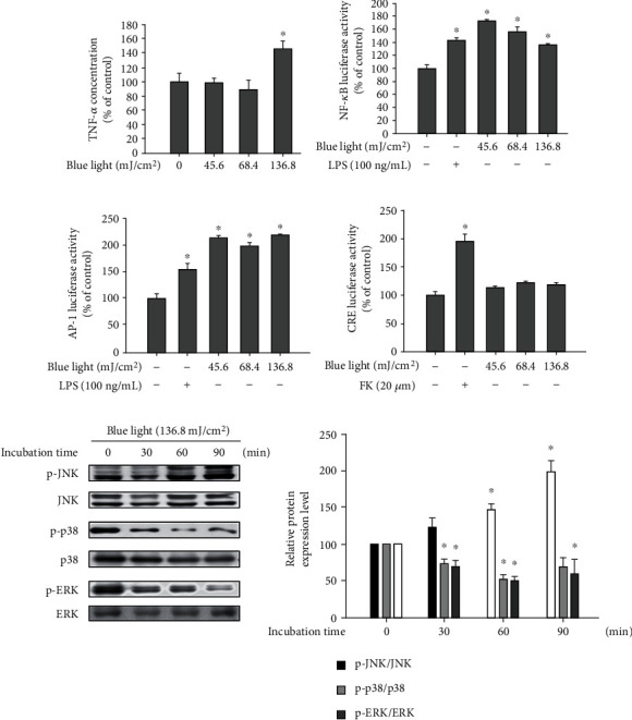
Figure 6.

Blue light upregulates proinflammatory cytokine production via NF-κB and AP-1 activation. (a) HaCaT cells were irradiated using the indicated intensity of blue light with a wavelength of 470–480 nm once daily for three consecutive days. On the fourth day, 24 h after the last irradiation, the cells were harvested and subjected to ELISA for analyzing the TNF-α levels. Data are presented as the mean ± SEM of four independent experiments. Statistical significance of differences among the groups was assessed by one-way analysis of variance (ANOVA), followed by Tukey's multiple comparison test, using the GraphPad Prism 5 software. ∗p < 0.05 vs. the control group. (b–d) HaCaT cells were cotransfected with the NF-κB, AP-1, or CRE promoter-luciferase reporters and β-galactosidase reporter vector using polyethylenimine. After 24 h, the transfected cells were irradiated with the indicated intensity of blue light. Twenty-four hours after the irradiation, the cells were harvested and subjected to luciferase reporter assay. Data are presented as the mean ± SEM of four independent experiments. Statistical significance of differences among the groups was assessed by one-way analysis of variance (ANOVA), followed by Tukey's multiple comparison test, using the GraphPad Prism 5 software. ∗p < 0.05 vs. the control group. (e) Cells were irradiated with blue light and incubated for the indicated time periods. The cells were harvested immediately after the incubation, and the protein levels of MAPKs and their phosphorylated forms were detected by Western blotting analysis.
