Figure 2.
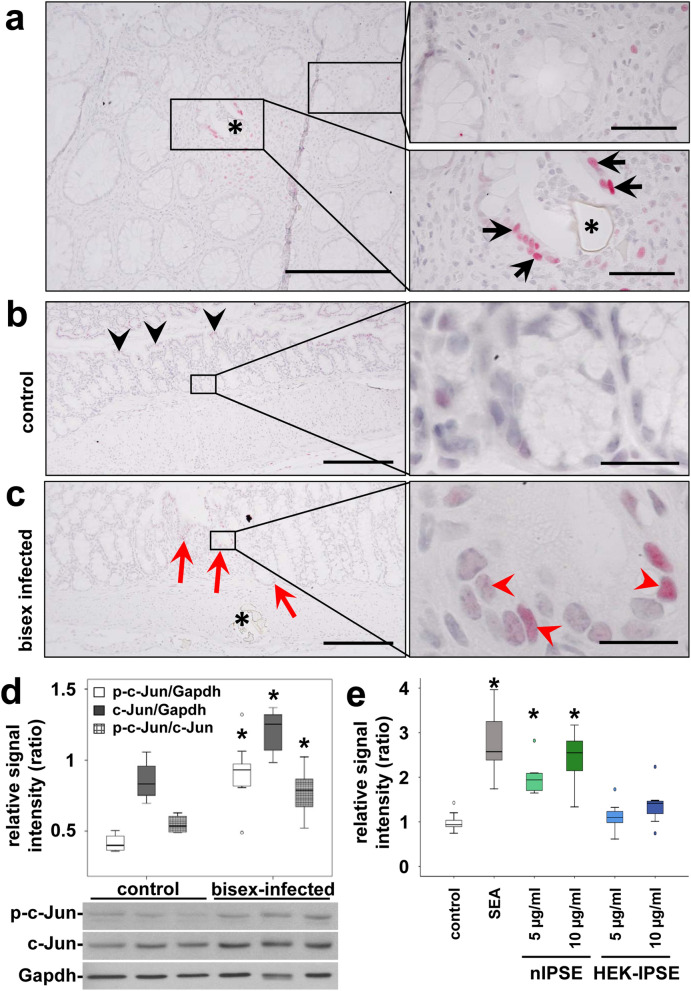
Activation of the protooncogene c-Jun in enterocytes at extravasation sites of S. mansoni eggs. (a) A representative histologic slice of a rectal biopsy from an 29 year-old Egyptian male with schistosomal colitis is shown. Immunohistochemical staining of c-Jun (red) depicted its nuclear translocation (arrows) in enterocytes of crypts in direct vicinity of S. mansoni eggs (*, lower right panel). Nearly no nuclear translocation of c-Jun was observed in enterocytes lining unaffected crypts at a distance of at least 200 µm (upper right panel). The same result was shown in at least three samples from different patients. Co-staining of nuclei in blue, magnification 200× and 1000×, bars 200 µm (left panel) and 50 µm (panel on the right). (b) Immunostaining visualized low amounts of epithelial c-Jun at the luminal side of the colon (black arrowheads), but not in enterocytes inside the crypts (magnification of the boxed area is depicted in the right panel). (c) Immunostaining demonstrated enhanced nuclear translocation of c-Jun (red arrowheads) in enterocytes of crypts (red arrows) adjacent to sites of S. mansoni egg (*) extravasation in bisex-infected hamsters. The quantification is presented in Suppl. Fig. 5. Magnified representative crypts are shown on the right. Magnification in (b,c) 200× and 1000×, bars 200 µm and 20 µm. (d) Western blot analysis and subsequent semi-quantitative assessment of optical density suggested an enhanced expression and activation of c-Jun (p-Ser73) in the colon of S. mansoni bisex-infected hamsters in comparison to single sex-infected hamsters, lacking egg production. The experiment and assay were reproduced at least two times. Representative blots are shown, control n = 4, bisex infection n = 11. Differences were analyzed statistically between control and bisex-infected for the activated form (p–c-Jun), total expression (c-Jun), and the ratio between p–c-Jun and c-Jun. *p ≤ 0.05. (e) The reporter gene assay demonstrated, that SEA and IPSE stimulation lead to the functional activation of the AP1 promotor. The experiment and assay were reproduced at least two times and analyzed by Kruskall-Wallis test. *p ≤ 0.05.

