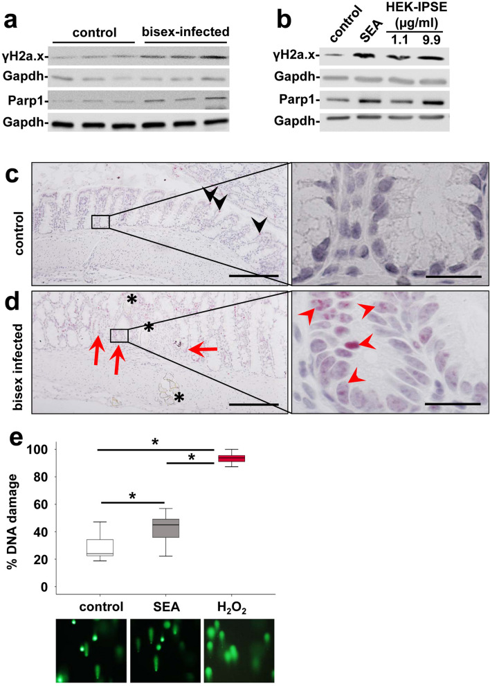
Figure 5.
S. mansoni egg-induced DNA repair. (a) Western blot analysis demonstrated enhanced expression of γH2a.x and Parp-1 in the colon of S. mansoni bisex-infected hamsters in comparison to single sex-infected controls. Representative blots are shown. (b) Induction of expression and activation of γH2a.x and Parp1 by SEA and HEK-IPSE is shown by western blot (stimulation 4 h). (c,d) Immunostaining of γH2a.x (blue) depicted nuclear staining (red arrows and arrowheads) in enterocytes of crypts in direct vicinity of S. mansoni eggs (*). Representative histologic section of the colon of a control hamster (c) and a bisex-infected hamster (d) are shown. Quantification of nuclear γH2a.x is demonstrated in Suppl. Fig. 10. Nuclear staining of γH2a.x in red, magnification 200×, bar 200 µm and 1000×, bar 20 µm. (e) The comet assay demonstrated enhanced DNA damage by SEA stimulation in SW620 cells. western blot.

