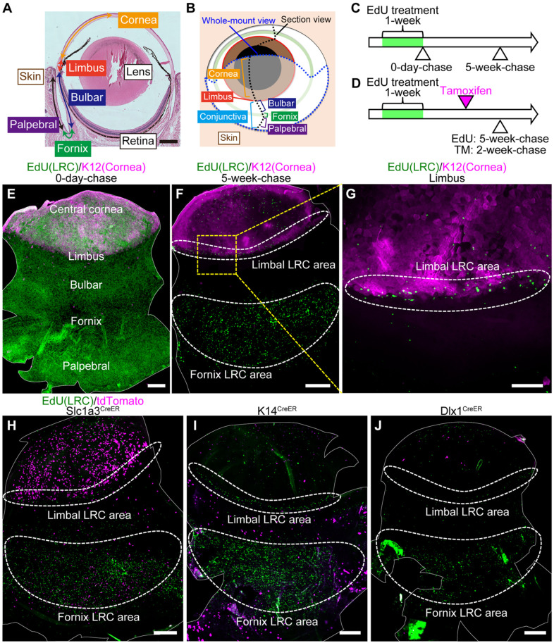
Fig. 1.
Distinct CreER tools mark label-retaining cell (LRC) and non-LRC compartments. (A) Hematoxylin-eosin stained mouse eye. (B) A schematic representation of the mouse eye. The eye was analyzed by sagittal sections (black dotted line) or whole-mount preparation of epithelial sheets (blue dotted line). (C) EdU pulse-chase scheme to detect LRCs in the ocular surface epithelium. (D) Scheme to examine the relationship of CreER+ cells with LRCs and non-LRCs. (E-G) Whole-mount staining of epithelial sheets after EdU pulse-chase experiments at 0-day-chase (E) and 5-week-chase (F,G). The solid white line outlines the whole-mount epithelial sheets (E,F). Limbal areas, surrounded by the yellow dashed square, are shown at a higher magnification in G. The white dashed line surrounds limbal and fornix LRC area. Green, EdU. Magenta, K12 (corneal marker). (H-J) Whole-mount staining of epithelial sheets after tamoxifen injection and EdU pulse-chase in Slc1a3CreER, K14CreER, and Dlx1CreER. The solid white line outlines the whole-mount epithelial sheets. The dashed white line surrounds the limbal and fornix LRC area. Green, EdU. Magenta, tdTomato. Scale bars: 500 μm (A,E,F,H-J); 200 μm (G).

