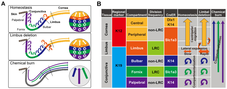
Fig. 6.
Proposed model of compartmentalized stem and progenitor populations with distinct cell division dynamics in the ocular surface epithelium. (A) Diagram representing the compartmentalization of the ocular surface epithelium and SC dynamics in homeostatic and post-injury conditions. (B) Summary table of genetic markers to define distinct SC compartments and lineage relationships. The long arrows represent the migration of cells from one compartment to another. The round arrows represent self-maintenance of each compartment by local SCs.

