Fig. 1.
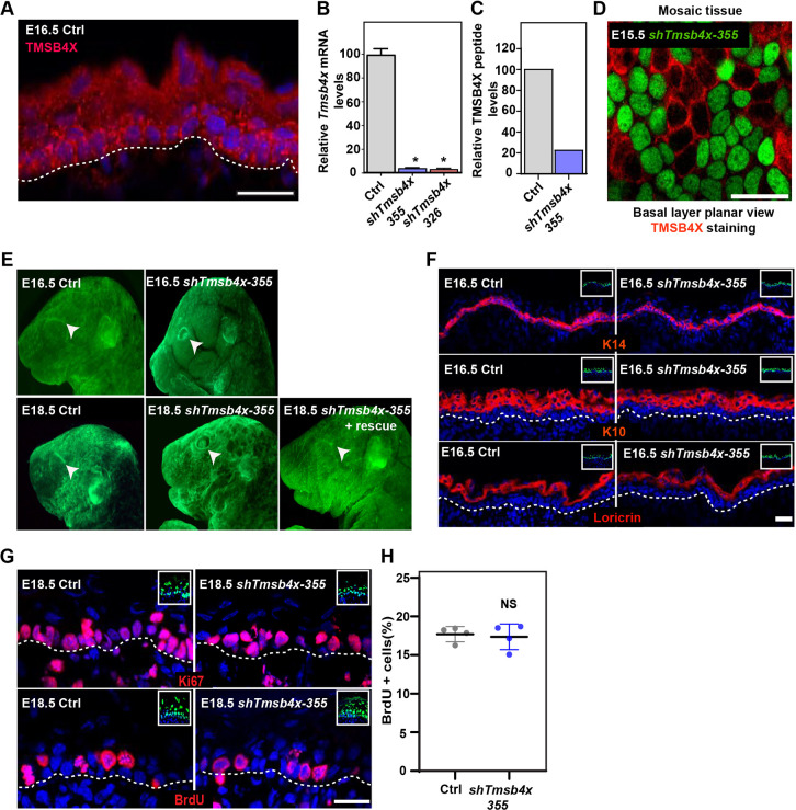
Tmsb4x depletion in the developing epidermis hinders eyelid closure. (A) Sagittal views of 10-μm sections of dorsal skin from E16.5 CD1 mouse embryo immunostained for TMSB4X. (B) RT-qPCR analysis of Tmsb4x mRNA levels in primary mouse keratinocytes transduced with scrambled shRNA (shScr; Ctrl) or one of two Tmsb4x shRNAs (326 or 355). Data are the mean±s.d. of five experiments. *P<0.0001 for Ctrl versus shTmsb4x-326 or shTmsb4x-355 by unpaired two-tailed t-test. (C) Liquid chromatographic analysis of three peptides derived from trypsin-digested TMSB4X isolated from shScr- (Ctrl) and shTmsb4x-expressing primary mouse keratinocytes. (D) Whole-mount immunofluorescence image of dorsal skin from E15.5 CD1 embryo injected on E9 with a Tmsb4x-355;H2B-GFP lentivirus and immunostained for TMSB4X. (E) Stereomicroscopic images of E16.5 and E18.5 embryos infected on E9 with shScr (Ctrl), shTmsb4x-355 or shTmsb4x-355+rescue lentiviruses. Arrowheads indicate open/closed eyes. (F) Sagittal views of 10-μm sections of dorsal skin from control (Ctrl) and shTmsb4x-355-transduced E16.5 embryos. Sections were immunostained for the basal layer marker K14, the differentiation marker K10 and the granular layer marker loricrin. (G) Sagittal views of 10-μm sections of dorsal skin from control and shTmsb4x-355-transduced E18.5 embryos. Sections were immunostained for the cell proliferation markers Ki67 and BrdU. (H) Quantification of BrdU+ cells from the data shown in E. Horizontal bars represent mean±s.d. from four embryos per condition, circles represent individual embryos. NS, not significant (P=0.3384) by unpaired two-tailed t-test. Nuclei were stained with DAPI (blue), dashed lines indicate the dermal-epidermal border, and insets show the transduced cells (H2B-GFP+). Scale bars: 20 μm.

