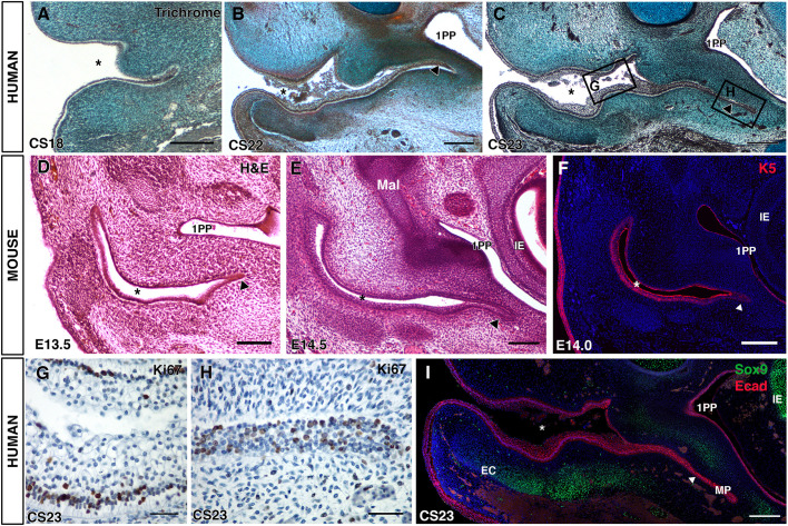
Fig. 1.
The ear canal forms in two distinct parts. (A-C) Trichrome-stained frontal sections through the developing human ear canal. (A) CS18, the meatus is just visible at the end of the open primary canal (asterisk). (B) CS22, the meatus (arrowhead) has extended towards the middle ear and first pharyngeal pouch (1PP). (C) CS23, the meatus has extended adjacent to the pharyngeal pouch as a thin sheet (arrowhead). (D-F) Forming and extending meatal plug, mouse frontal sections stained with Eosin (D,E) and for keratin 5 (F). (D) At E13.5 the meatal plug (arrowhead) is only evident as a small group of cells at the end of the open primary canal. (E) At E14.5, the meatal plug has extended towards the middle ear (arrowhead). (F) Keratin 5 expression at E14.0 in the primary canal (asterisk) and meatus (arrowhead). (G,H) Ki67 in human frontal sections. Images show regions indicated by rectangles in C. (G) The open primary canal with positive (brown) cells located at the basal layer. (H) The meatal plug with positive (brown) cells throughout the epithelium. (I) CS23, immunofluorescence for Sox9 (green) and E-cadherin (pink). DAPI nuclear stain (blue). The two parts of the canal [primary canal and meatal plate (MP)] are clearly visible. The developing external ear canal cartilages (ECs) are observed forming under the primary canal but not the meatal plate. Asterisks in A-F,I indicate the open primary canal. Scale bars: 500 µm in A-C; 200 µm in D-F,I; 100 µm in G,H. 1PP, first pharyngeal pouch; IE, inner ear; EC, ear canal cartilage; MP, meatal plate.

