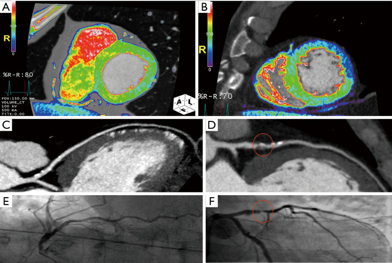
Figure 4.
First-pass myocardial perfusion imaging evaluation during angiographic coronary evaluation (curved-MPR LAD image) and ICA control. Normal LV first pass perfusion related to calcific non obstructive CAD of LAD (A, CCTA short axis color map; C, LAD curved-MPR; E, ICA) and perfusion defect of the anterior and anterolateral LV wall related to LAD obstructive mixed coronary plaque with ICA control (B, CCTA short axis color map; D, LAD curved-MPR; F, ICA). LAD, left anterior descending coronary artery; CAD, coronary artery disease; ICA, invasive coronary angiography; red circle, proximal LAD severe obstructive coronary plaque.

