
Fig. 5.
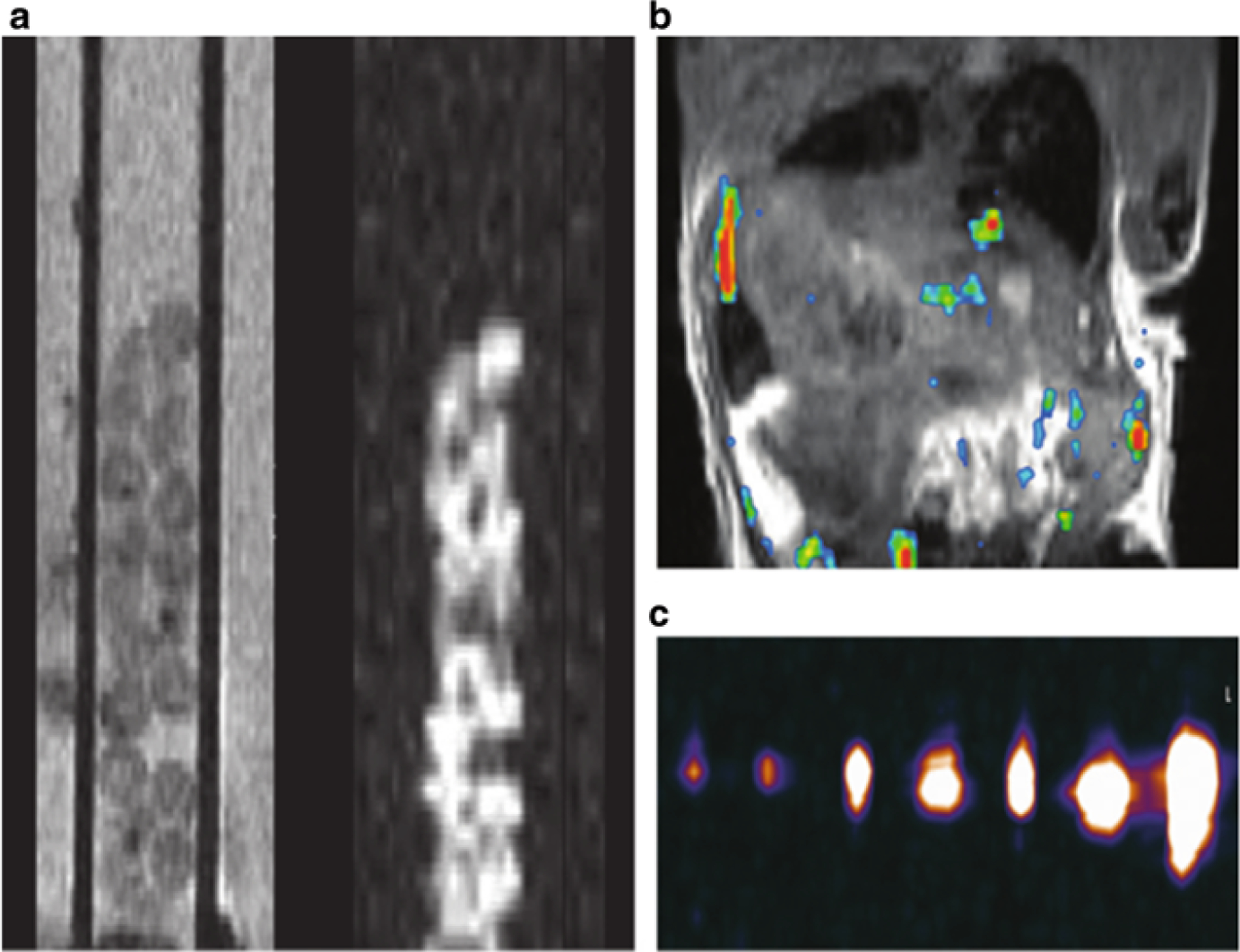
19F MR-trackable capsules. (a) In vitro proton T2-weighted MR image (left) and corresponding 19 F MR image (right) of fluorocapsules in a standard 5 mm glass tube, obtained with a 11.7 T preclinical system. (The capsules were introduced in a 500 mm capillary tube.) (b) In vivo 19 F MR image of a mouse after intraperitoneal transplantation of 2000 fluorocapsules. An overlay of the 19 F image (pseudocolor) on the 1H image (gray scale) is shown. (c) Fluorine 19 MR image of fluorocapsules obtained with a 3.0 T clinical system. From left to right, images are of gel phantoms containing 1, 2, 5, 10, 15, 25, and 50 capsules. Reprinted with permission from ref. 35. Copyright © 2015 Radiological Society of North America