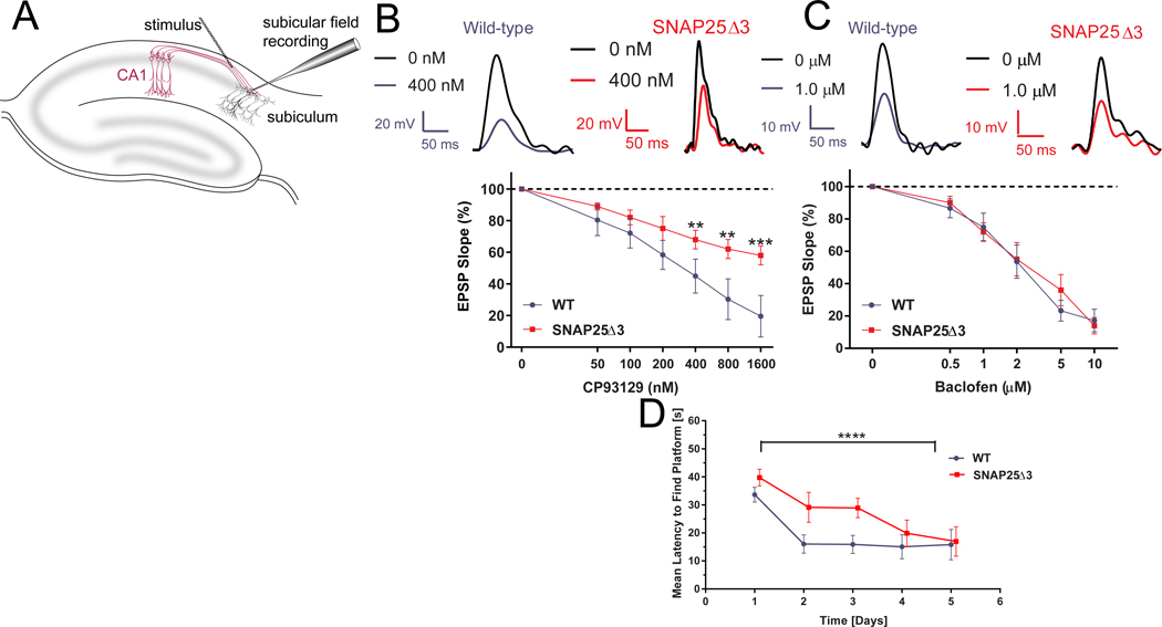
Figure 4. SNAP25Δ3 homozygotes have impaired Gi/o-coupled GPCR signaling in CA1/subiculuar hippocampal neurons and show impaired hippocampal spatial learning.
A. Diagram of the hippocampal field recording paradigm. Stimulation with bipolar electrodes over the CA1-subicular pathway evoked field EPSPs recorded in basal dendrites of subicular pyramidal neurons in AP5 (50 μM) and bicuculline (5μM) to isolate AMPAR-mediated responses. B. Traces from CA1-subicular recordings in WT (left panel) and SNAP25Δ3 (right panel) slices at 0 and 400 nM CP93129. Bottom panel: Dose response of the effect of CP93129 on the AMPA component of these field EPSPs from 6-week old littermate WT (in blue) or SNAP25Δ3 homozygotes (in red). Amplitudes were normalized to the control response. CP93129 was significantly more potent in WT than SNAP25Δ3 (**p = 0.0068, 400 nM; **p = 0.0035, 800 nM; ***p = 0.001, 1600 nM; Student’s t-test). WT: n= 8 slices from 6 mice. SNAP25Δ3: n= 5 slices from 5 mice. C. Traces from field recordings in wild-type (left panel) and SNAP25Δ3 (right panel) slices at 0 and 1.0 μM baclofen. Bottom panel: Dose-response of the effect of baclofen on field EPSPs recorded in the WT or SNAP25Δ3 hippocampal slices. No significant differences were detected by genotype. WT and SNAP25Δ3: n= 5 slices from 5 mice for both genotypes. D. Comparison between age-matched littermate WT and SNAP25Δ3 homozygotes in the acquisition of the Morris Water Maze Task over a 5 day trial period by genotype (p<0.05) and time (p<0.0001). n=11 WT mice and 11 SNAP25Δ3 mice.

