Figure 5.
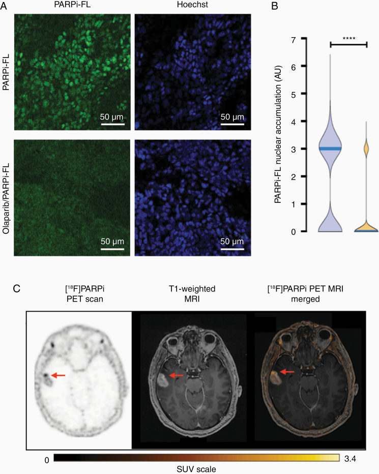
Biospecimen and imaging of lesion #7, untreated glioblastoma. (A) PARPi-FL uptake blocking demonstrates the specificity of the compound. Biospecimen stained with the fluorescent version (PARPi-FL, top row) and blocked (co-incubated with a 100-fold excess of olaparib, bottom row). (B) Quantification of nuclear accumulation of PARPi-FL showed median fluorescence significantly higher (P < .001) than in the blocked tissue. (C) Axial [18F]PARPi uptake map, contrast T1-weighted image, and [18F]PARPi map overlaid on contrast T1-weighted image show untreated enhancing cancer in the temporal lobe with high [18F]PARPi uptake (arrow).

