
Figure 2. Examples of erythema migrans (EM).
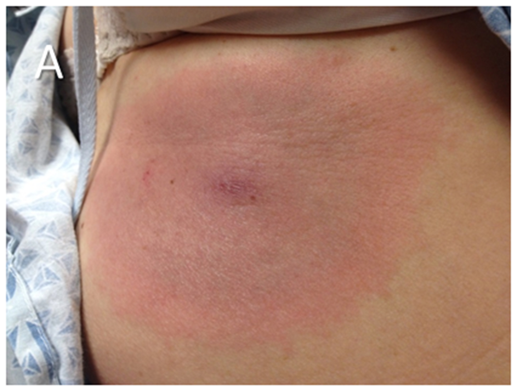
(A) An EM skin lesion that developed at the site of a tick bite on the abdomen of a patient. The lesion is circular and homogeneous, a pattern which is more common than the well-recognized “bull’s eye” appearance of the skin lesion. The primary EM lesion typically is at least 5 cm in diameter. (B) Multiple EM lesions on the back of a patient during disseminated Lyme disease. Secondary EM lesions may be smaller than the primary lesion. The photograph in panel A is courtesy of Dr. Roger Clark, Faulkner Hospital, Boston, MA.