
Figure 4. Babesia microti parasites in human red blood cells.
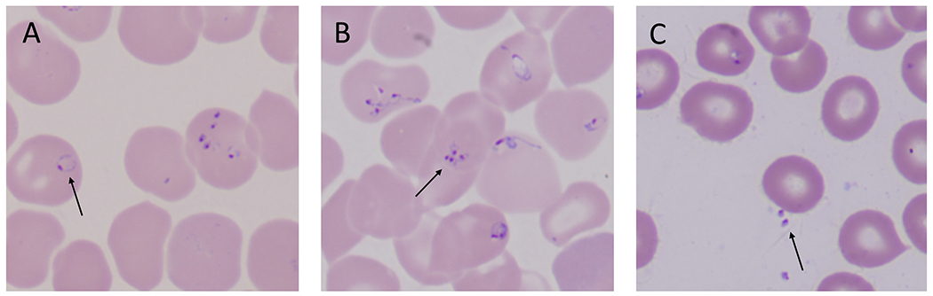
(A) B. microti trophozoites often appear as rings with one chromatin dot. Arrow points to a classic ring form of babesia. (B) Asexual division of the parasite yields up to four merozoites which can arrange in a tetrad, also known as Maltese cross (see arrow). Maltese crosses can be formed by B. microti, B. duncani, and B. divergens in human red blood cells. (C) Following rupture of an infected red blood cell, free merozoites (see arrow) quickly seek to adhere and invade an intact red blood cell. Micrographs are courtesy of Rouette Hunter and Stephen Johnson from the Hematology Laboratory at Tufts Medical Center, Boston, MA.