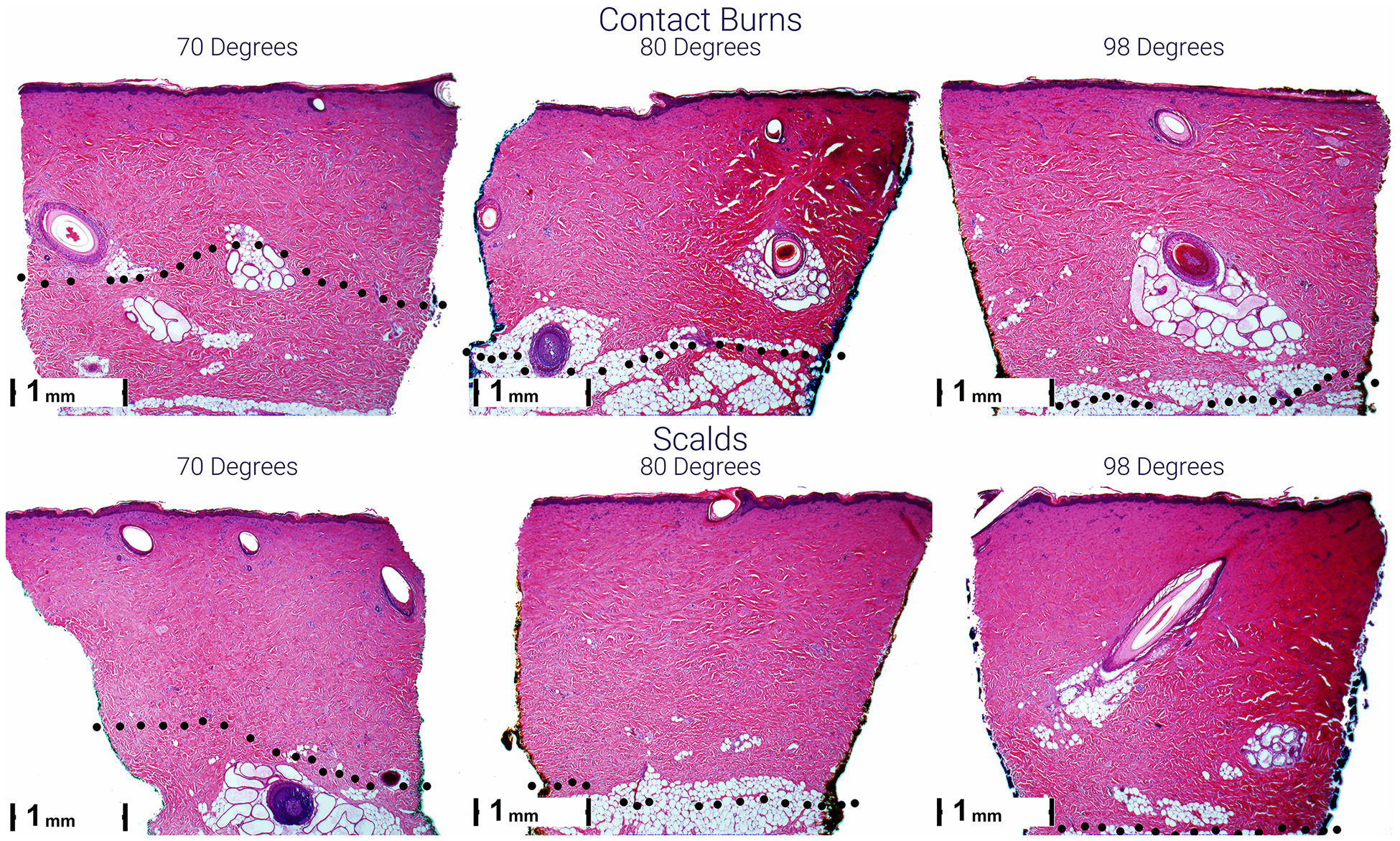
Figure 3.

Representative micrographs of scald (lower) and contact (upper) burns 1 hour after injury. Burn depth is greater from left to right and top to bottom. The black dots indicate the lower boundary of the burn with evidence of blood vessel occlusion and/or necrosis of endothelial cells and hair follicle and sebaceous gland cells.
