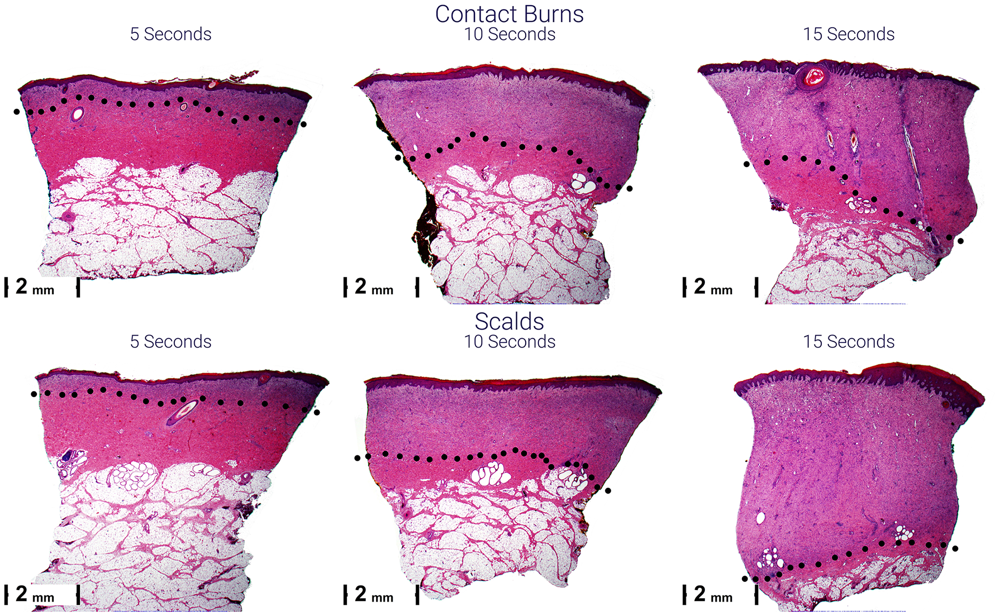
Figure 4.

Representative micrographs of scald (lower) and contact (upper) burns 28 days after injury. Scars become thicker from left to right and top to bottom. The black dots indicate the lower boundary of the scar. The purple scars are due to the cellular infiltrate of granulation tissue composed of plump fibroblasts and new vessels. This is compared to the normal pink staining in the dermis below.
