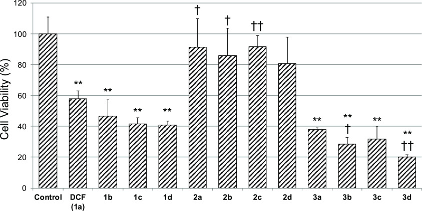
Figure 1.
Cytotoxicity of DCF and its analogs in cryopreserved human hepatocytes. Each bar shows the cell viability after incubating with test compounds (200 μM) for 2 h with shaking. Cell viability was assessed by WST-8 assay, and the data are expressed relative to the control group. The control group was treated with 0.25 v/v% DMSO. Each value represents the mean ± S.D. of three samples. **p < 0.01; significantly different from control, ††p < 0.01, †p < 0.05; significantly different from DCF, Dunnett’s post hoc test.

