
Figure 1. Eye development in Xenopus laevis.
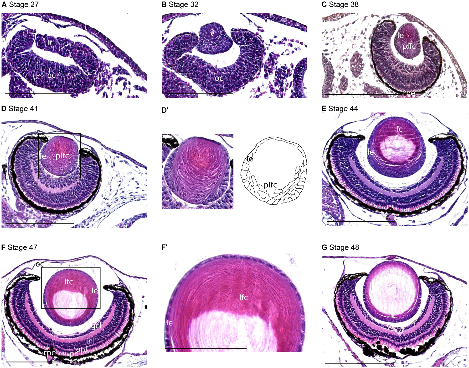
Representative sections of embryos staged according to Nieuwkoop and Faber58, hematoxyline-eosine stained. A, Stage 27, 30 hours post-fertilization at 23°C. lr lens rudiment; oc optic cup. B, Stage 32, 40 hours post-fertilization. lv lens vesicle. C, Stage 38, 2 days 5 hours post-fertilization. le lens epithelium; plfc primary lens fiber cells; rpe retinal pigmented epithelium. D, Stage 41, 3 days 4 hours post-fertilization. D’, Higher magnification of the lens shown in D, with interpretative diagram. E, Stage 44, 3 days 20 hours post-fertilization. lfc, lens fiber cells. F, Stage 47, 5.5 days post-fertilization. gcl, ganglion cell layer; inl, inner nuclear layer; oc, outer cornea; onl, outer nuclear layer; pr, photoreceptors; rpe retinal pigmented epithelium. F’, Higher magnification of the lens shown in F. G, Stage 48, 1 week post-fertilization. All scale bars 200 μm except F’ 90 μm.