
Figure 4. Eye phenotype of dnase2b crispants.
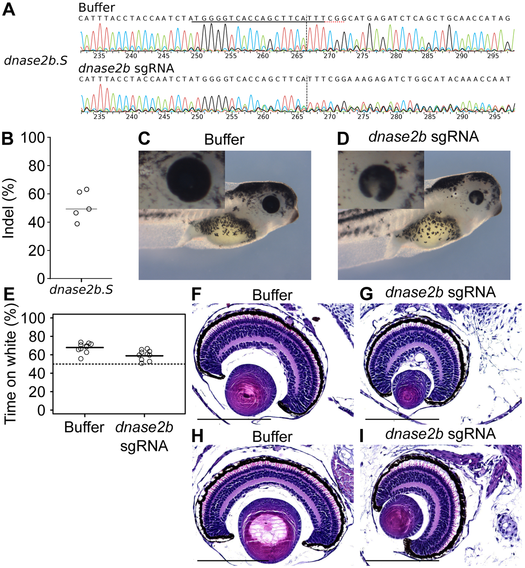
We injected embryos with Cas9 enzyme and a mixture of sgRNA targeted against dnase2b.S and a putative dnase2b.L locus, and we allowed the embryos to develop. A-B, Representative Sanger chromatograms of one sgRNA and one buffer-injected embryo and percentages of InDels in individual embryos, as in Figures 2A–B. Since we failed to amplify the putative dnase2b.L locus, only the results for dnase2b.S are shown. C-D, Stage 41 larvae previously injected with buffer (C) or Cas9-sgRNA (D). Insets are high magnifications of the eyes of the same larvae. E, Functional vision assay of buffer-injected embryos and dnase2b crispants, as in Figure 2F. F, Histological section of the eye of a buffer-injected stage 41 tadpole. G, Section of the eye of a Cas9 and sgRNA-injected stage 41 tadpole with microphthalia. H-I, Same as F-G with stage 45 tadpoles. Scale bars 200 μm.