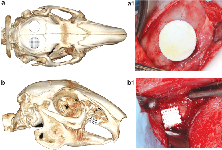
FIG. 4.
micro-CT image of rabbit skull, schematic depicting (a) calvarial defect and (b) alveolar cleft model, respectively. The skeletally immature rabbits underwent surgical resection of alveolar calvaria and alveolar cleft, each site receiving custom-designed and 3D printed β-TCP scaffolds. Intraoperative placement of scaffolds in (a.1) calvaria and (b.1) alveolar ridge defects using a fit-and-fill process.

