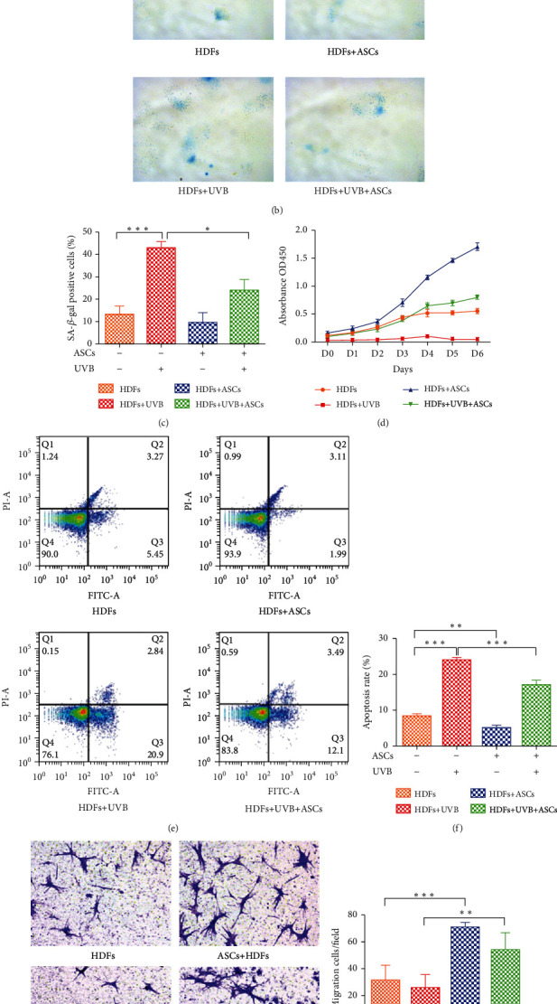
Figure 1.

Assessment of the functions of HDFs cocultured with ASCs. (a) Schematic illustration of the cell experiment. (b) Representative images of senescence-associated β-galactosidase (SA-β-gal) staining. Positive blue staining of SA-β-gal appeared in senescent HDFs. (c) The average percentage of SA-β-gal-positive cells was quantified. Five random fields were selected for counting. (d) Proliferation of HDFs was assessed by CCK-8 assays. (e) HDF apoptosis was measured by FACS analysis after Annexin V-FITC/PI staining. (f) The apoptosis rates of HDFs were quantified. (g) Representative images of migrated HDFs (magnification, ×200). (h) The number of migrated HDFs per field. Five random fields were selected for counting. All experiments were independently conducted in triplicate, and the values are expressed as the mean ± SD. (∗p < 0.05, ∗∗p < 0.01, ∗∗∗p < 0.001).
