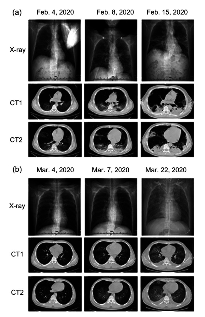
Fig. 1.
Comparisons of chest imaging (X-ray and CT scans) of COVID-19 and CMV pneumonia
These two types of pneumonia (COVID-19 vs. CMV) showed a somewhat similar progression in the initial stage, progressive stage, and consolidation stage/diffuse infiltration stage of infection. Initially, both presented with typical GGOs. (a) Chest imaging of COVID-19 pneumonia. A 51-year-old male was confirmed with COVID-19 and received CT scans on 4, 8, and 15 February, 2020. (b) Chest imaging of CMV pneumonia. A 26-year-old male with diffuse large B cell lymphoma (non-GCB, IVB stage) was diagnosed with CMV pneumonia during chemotherapy. He received CT scans on 4, 7, 22 March, 2020. COVID-19, coronavirus disease 2019; CMV, cytomegalovirus; CT, computed tomography; GCB, germinal center B-cell-like lymphoma; GGOs, ground-glass opacities

