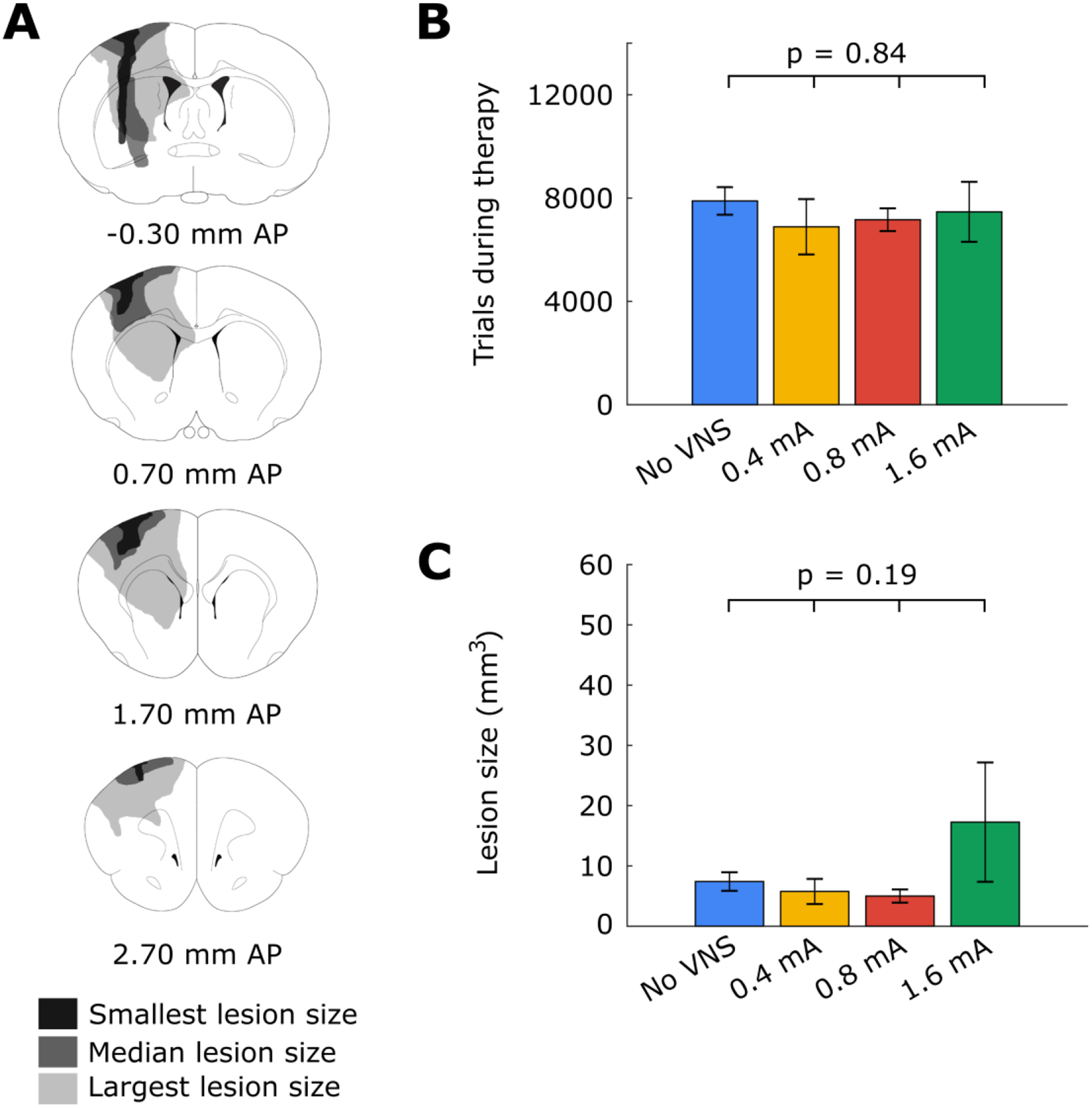
Figure 3:

(A) Illustrated coronal sections demonstrating the smallest, median, and largest motor cortex lesions observed in the study. (B) There was no significant difference in the number of trials initiated across all experimental groups. (C) There was no significant difference in lesion volume across all experimental groups. Error bars represent SEM.
