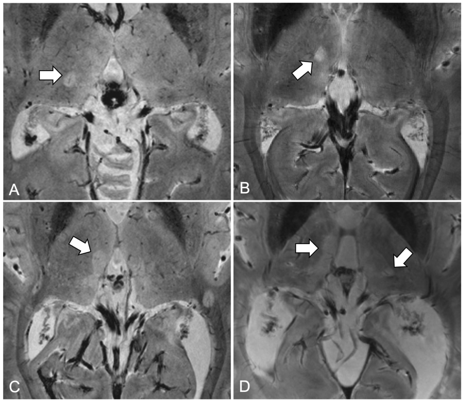
Figure 2. Examples of thalamic multiple sclerosis lesions on 7 Tesla T2*-weighted MRI images.

(A) Ovoid lesion with central venule on right thalamus; (B) Ovoid lesion without central venule on right thalamus; (C) Periventricular lesion on third ventricle; (D) Multiple thalamic lesions including periventricular and ovoid lesions.
