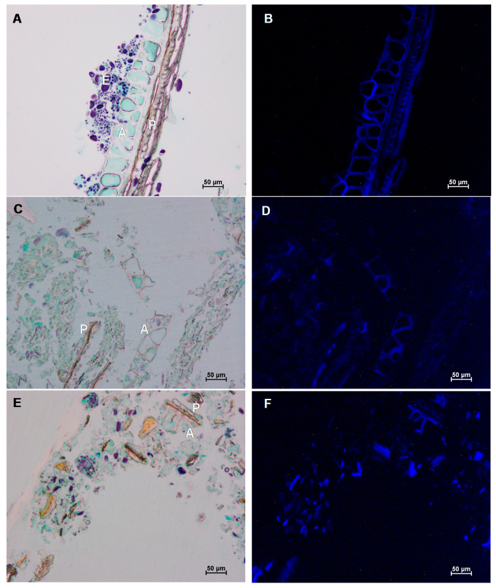
Figure 3.
Visualisation of untreated coarse wheat bran (A,B) and wheat bran that was cryogenically milled (C,D) or wet milled (E,F) on a large scale. In the milled samples almost all bran structures were broken down but in these pictures we focused on a few partially intact structures. Samples were stained with Lugol, Light Green and Calcofluor and visualised with light microscopy (A,C,E) and fluorescence microscopy (B,D,F). The pericarp (P), aleurone (A) and residual endosperm (E) are indicated.

