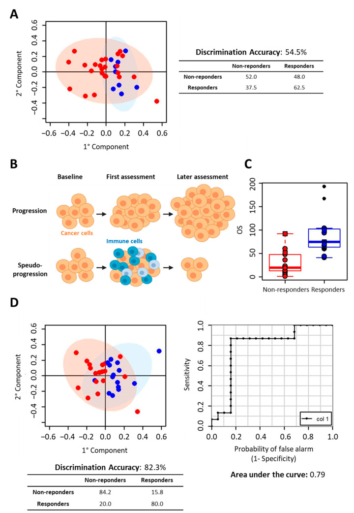
Figure 2.
Prediction of Nivolumab response. (A) Discrimination between responders and non-responders according to the first radiological assessment. O-PLS analysis of T0 samples. Score plot, PC1 vs. PC2, and corresponding confusion matrix. Red dots: non-responder subjects; blues dots: responder subjects. (B) Scheme of disease progression vs. pseudo-progression in the presence of immune cells attacking the tumour. (C) Boxplots of OS values of the subject. Red dots: non-responder subjects; blue dots: responder subjects (subjects still on alive are marked with black dots). (D) Discrimination between responders and non-responders according to the second radiological assessment. O-PLS analysis of T0 serum samples. Score plot, PC1 vs. PC2, and corresponding confusion matrix (left panel); ROC curve derived from O-PLS cross-validation (right panel). Red dots: non-responder subjects; blues dots: responder subjects.

