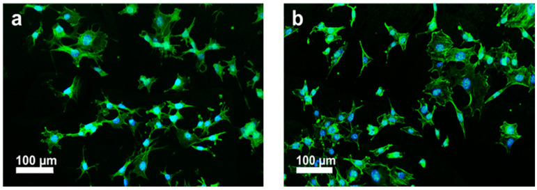
Figure 25.
Fluorescence micrographs of murine MC3T3-E1 pre-osteoblasts cultivated on (a) PEEK/1.5 wt% MWNT and (b) PEEK/3.0 wt% MWNT nanocomposites for 3 days, and stained with F-actin cytoskeleton (green) and nucleus (blue). Reproduced from [63] with permission of MDPI.

