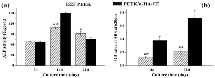
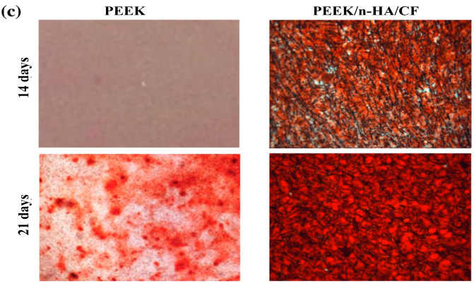
Figure 27.
(a) ALP activity normalized to protein content of MG63 cells cultured on pure PEEK and PEEK/nHA-SCF hybrid for different time points. (b) The quantitative value of matrix mineralization acquired by measuring optical density. (c) Alizarin red staining for calcium-rich deposits secreted by MG63 cells on PEEK and PEEK/nHA-SCF hybrid. * represents p < 0.05 and ** indicates p < 0.01 compared with PEEK/nHA-SCF. Reproduced from [200] with permission of Elsevier.


