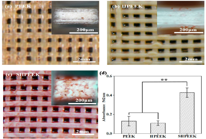
Figure 30.
Alizarin red S staining of calcified nodules secreted by MC3T3-E1 cells on different scaffolds at day 7. Optical photographs of Alizarin red S staining of MC3T3-E1 cells on (a) PEEK, (b) HPEEK, and (c) SHPEEK scaffolds. (d) Quantitative analysis of deposited calcified nodules. (** p < 0.01). Reproduced from [216] with permission of Elsevier under a Creative Commons license.

