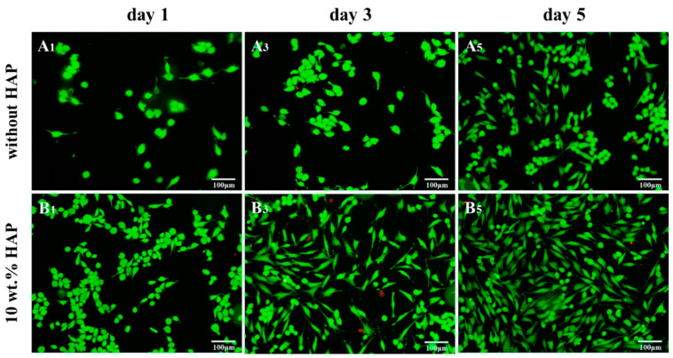
Figure 34.
Fluorescence images of MG63 cells cultured on PEEK-20 wt% PGA (top panel) and PEEK-20 wt%PGA/10 wt% nHA (bottom panel) scaffolds for different time periods. HAP: nanohydroxyapatite. Scale bar: 100 µm. Reproduced from [220] with permission of MDPI under the terms and conditions of the Creative Commons Attribution license.

