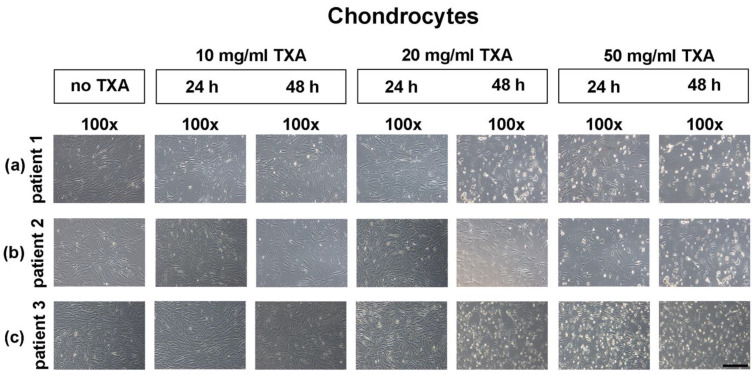
Figure 3.
Microscopical analysis of chondrocyte growth in monolayer cultures after treatment with different concentrations of tranexamic acid for varying exposure times. Chondrocytes were harvested from the hyaline hip cartilage of three separate patients that underwent total hip arthroplasty. Cells were seeded in cell culture flasks, incubated in cell culture medium and grown to confluency. After reaching confluency, controls were maintained (no TXA), while other cells were exposed to 10 mg/mL, 20 mg/mL or 50 mg/mL of TXA. The exposure time varied from 24 h to 48 h. Representative samples from three different donors were captured directly after treatment with TXA at low (100×; black bar = 200 μm) magnification. TXA, tranexamic acid; min, minutes; d, days; h, hours.

