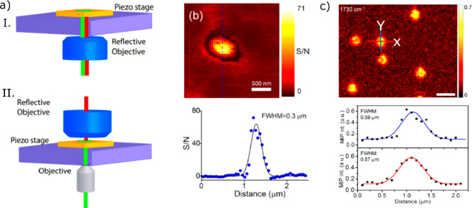
Figure 13.
Co-propagating (I) and counter-propagating (II) configurations of mid-IR photothermal microscopy. In the co-propagating scheme, the pump beam (red) and the probe beam (green) propagate in the same direction, whereas the pump and probe beams counter-propagate in the counter-propagation scheme. Adapted with permission from ref (120). Copyright 2020 Royal Society of Chemistry. (b) Mid-IR photothermal imaging of a 100 nm polystyrene bead (top) and the line profile (bottom) along the dashed line shown at the top. The full width at half-maximum (fwhm) is 300 nm. Adapted from ref (62). Copyright 2017 American Chemical Society. (c) Mid-IR photothermal imaging of 500 nm PMMA beads (top) and horizontal (red) and vertical (blue) line profiles (bottom). The fwhm along X and Y directions were 590 and 570 nm, respectively. The deconvolution of the image spot with the particle size resulted in a fwhm of about 290 nm. Scale bar: 1 μm. Adapted with from ref (68). Copyright 2019 American Chemical Society.

