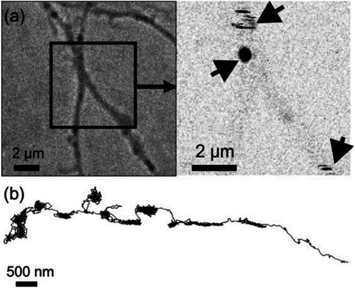
Figure 17.
(a) White-light image (left) and photothermal image (right) of a live neuron labeled with gold nanoparticles marked with arrows. A photothermal image of gold nanoparticles shows a nice alignment with the white-light image of the live neuron. The photothermal image shows both solid and stripe patterns. The solid pattern is due to slow, confined diffusion, whereas the striped pattern is due to fast diffusion of the nanoparticle. (b) Trajectory of a 5 nm gold nanoparticle in a live neuron recorded at a video rate, showing the heterogeneity in its diffusion. The particle’s movement alternates between slow and fast diffusion. Reprinted with permission from ref (149). Copyright 2006 Elsevier.

