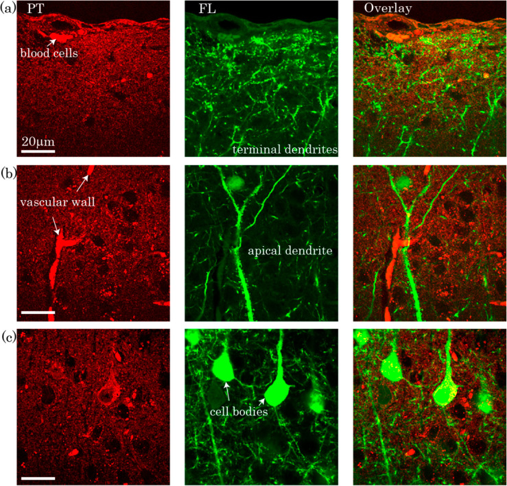
Figure 18.
Combined photothermal (left), fluorescence (center) images, and their overlays (right) of different layers of mouse brain cortex at (a) dendrite terminals, (b) dendrite, and (c) blood cells. Both photothermal and fluorescence images represent neurons expressing yellow fluorescent proteins (YFP). Photothermal signals show more distinct structures and organizations producing strong photothermal signal at the red blood cells and vessels probably because they include heme proteins. A Y-shaped vessel is observed in the photothermal image shown in (b) possible due to red blood cells fragmenting and stacking inside the vessels. The pump and probe beam powers were 0.5 and 4 mW, respectively. The image acquisition time was 6 s for an area of 500 × 500 pixels. Reprinted with permission from ref (148). Copyright 2016 The Optical Society (OSA).

