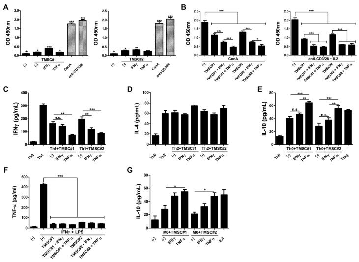
Figure 3.
Enhanced immunomodulatory potency of TNF-α-primed TMSCs. TMSCs isolated from two different individuals were cultured for 24 h in the absence or presence of either IFN-γ or TNF-α. (A) Naïve or primed TMSCs were cocultured with PBMCs without any stimulants to assess the immunogenicity and proliferation of PBMCs was determined using BrdU cell proliferation ELISA kit, compared to ConA- or anti-CD3/28-treated PBMCs as positive controls; (B) Naïve or primed TMSCs were cocultured with PBMCs in the presence of ConA or anti-CD3/28 plus IL-2 and the suppressive effects on the immune cell proliferation were determine; (C,D) Naïve or primed TMSCs were added in the process of Th cell differentiation into Th1 or Th2 subtype and production of typical cytokines, (C) IFN-γ or (D) IL-4, was measured in the cell culture supernatant; (E) Treg cell induction capability of naïve and primed TMSCs was evaluated through quantifying the IL-10 concentration in the coculture supernatant; (F,G) To assess the regulatory effects on macrophage polarization, naïve or primed TMSCs were cocultured with THP-1-derived macrophages; (F) Primed or unprimed control TMSCs were cocultured with classically activated M1 type macrophages and the secretion of TNF-α was measured in the conditioned media; (G) Naïve or primed TMSCs were cocultured with resting M0 macrophages for 2 days and the production of IL-10 was determined using ELISA to assess the anti-inflammatory M2 type macrophage induction potential. All data are represented as mean ± S.D. p-value = * < 0.05, ** < 0.01, *** < 0.001, compared to the control group marked as ‘+’.

