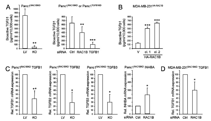
Figure 1.
RAC1B depletion controls TGFβ1 gene expression and secretion in Panc1 and MDA-MB-231 cells. (A) Concentration of bioactive TGFβ1 in culture supernatants, as measured by ELISA, of Panc1 cells genetically engineered to lack exon 3b of RAC1 and hence expression of RAC1B (Panc1RAC1BKO) or empty lentiviral vector (LV) control cells [7] (827.4 ± 166.9 vs. 35.2 ± 40.7, n = 4, p = 0.0012, left-hand graph), or PancRAC1BKD and ctrl cells (393.3 ± 98.2 vs. 264.8.2 ± 98.3, n = 3, p = 0.0038) or Panc1TGFB1KD and ctrl cells (393.3 ± 98.2 vs. 85.5 ± 38.9, n = 3, p = 0.029, right-hand graph). Cells were allowed to condition the media for 24 h. (B) As in (A), except that culture supernatants were retrieved from two individual clones (cl.) of MDA-MB-231 cells stably transfected with HA-RAC1B. Data shown are representative of three assays performed in total (means ± SD from triplicate samples). (C) Panc1RAC1BKO cells were subjected to qPCR analyses of TGFB1 (n = 5), TGFB2 (n = 3), or TGFB3 (n = 3), whereas Panc1RAC1BKD cells underwent qPCR analysis of INHBA. (D) MDA-MB-231RAC1BKD cells were subjected qPCR analysis of TGFB1. Data in (C,D) are the mean ± SD of three different transfections. The asterisks indicate significant differences (* p < 0.05; ** p < 0.01; *** p < 0.001). Successful KO or KD of RAC1B or TGFB1 in Panc1 and MDA-MB-231 cells, and verification of ectopic overexpression of HA-RAC1B in MDA-MB-231 cells is shown in Figure S1B.

