Figure 2.
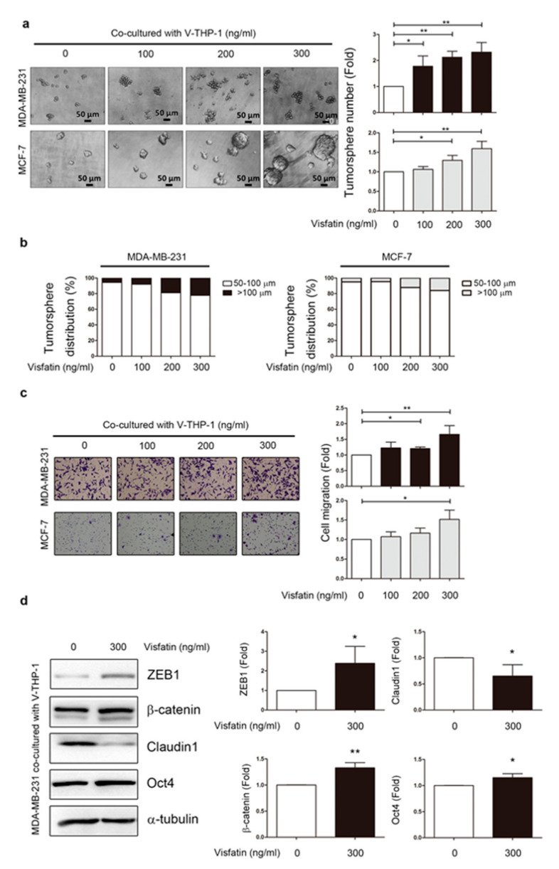
Visfatin-treated THP-1 enhanced tumor development in breast cancer cells. (a) MDA-MB-231 and MCF-7 were co-cultured with V-THP-1 at different doses (0, 100, 200, 300 ng/mL) for two days, and tumorsphere formation was observed by monitoring the size of the suspension cell clusters for another seven days. Cell clusters with size over 50 μm were counted as positive tumorsphere formation. Image scale bar, 50 μm. (b) Tumorsphere sizes defined as 50–100 μm and ≥100 μm were plotted in a bar chart. (c) MDA-MB-231 and MCF-7 cells were co-cultured with visfatin-treated THP-1 at different doses (0, 100, 200, 300 ng/mL) for 24 h, and migration ability was assessed in a trans-well system (40×). (d) MDA-MB-231 was co-cultured with visfatin-treated THP-1 (0, 300 ng/mL) for 24 h, and EMT and stemness markers were evaluated by Western blotting. Statistical analysis was done by the t-test. p-value ≤ 0.05 marked as *; p-value ≤ 0.01 marked as **.

