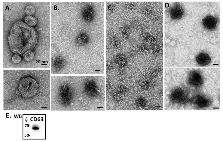
Figure 2.
Transmission electron microscopy (TEM) analysis of Mollusca hemolymph EVs. (A) Blue mussel; (B) soft shell clam; (C) Eastern oyster; (D) Atlantic jacknife clam. (E) Western blotting (WB) of hemolymph EVs (representative figure showing EVs from soft Atlantic jacknife clam) shows strong CD63 positive (protein size standard is indicated in kilodaltons, kDa).

